
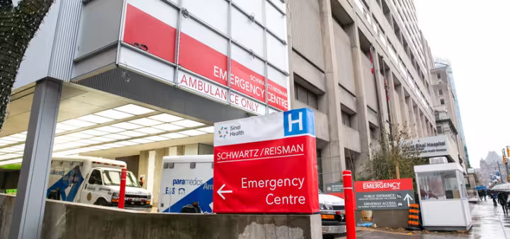
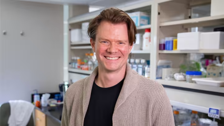


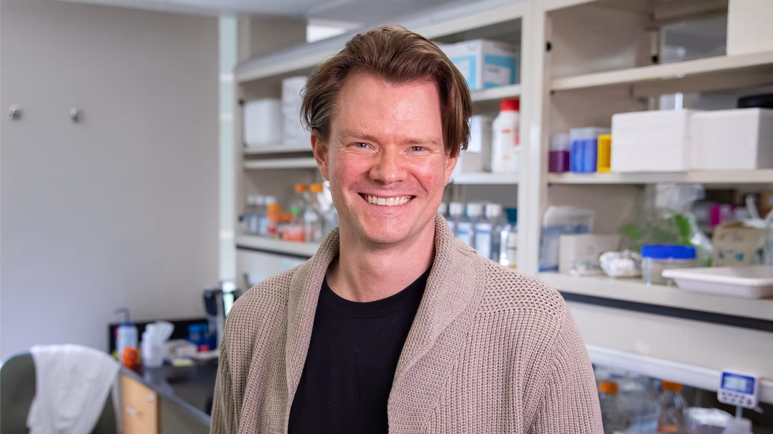
Meet the Minds: How Dr. Hartland Jackson is mapping the hidden complexity of cancer, one cell at a time
Cancer is often thought of as a single disease or a single mass of abnormal cells. But inside every tumour is a complex ecosystem – different cell populations interacting, adapting and responding to their environment in different ways. Understanding that complexity may be key to improving how cancer is diagnosed and treated.
Dr. Hartland Jackson – an Investigator at the Lunenfeld-Tanenbaum Research Institute (LTRI) and Associate Scientist at Ontario Institute for Cancer Research – studies how cells organize themselves within healthy tissues and how that organization breaks down in diseases like pancreatic and breast cancer. Using advanced imaging technologies and computational analysis, his research aims to map tumours at the cellular level to help drive more precise, personalized cancer care.
Q. How would you describe what you study in the simplest terms?
Dr. Jackson: At its core, my team and I are trying to understand how cells organize themselves – both in healthy tissues and in disease. Healthy cells communicate and work together to form structured, functional tissues. But in cancer, that organization breaks down. Cells stop following the rules, and instead of cooperating, they grow in a disorganized way that forms tumours.
Q. What does the difference between healthy and cancerous tissue look like?
Dr. Jackson: Healthy tissues are beautifully organized. They're structured for a purpose, whether that looks like tubes to secrete milk in breast tissue or in the shape of sacs to produce digestive enzymes in pancreatic tissue. In cancer, these structures are destroyed. Tumour cells grow everywhere and into surrounding tissues, and they compete with healthy cells for space and resources. So, it’s really the loss of organization that stands out. In fact, that’s how pathologists diagnose cancer. They’ll look at images of the tumour and ask: how far has this tissue moved away from normal organization?
Q. You study cancer at the single-cell level. Why is this so important?
Dr. Jackson: Individual cells are the fundamental building blocks of a tumour – almost like the people that make up a community. On their own, each cell is making decisions and carrying out specific functions, but together they communicate and organize into something larger. In healthy tissue, that collective behaviour creates functional structures that do important jobs, like producing milk or digestive enzymes. In cancer, those same systems break down, and the “community” begins behaving differently – growing, invading surrounding areas and doing harm.
To truly understand cancer, you have to look at both levels at once: the individual cells and the bigger picture they create together. A cell’s behaviour is shaped not only by its own internal decisions, but also by its environment and the local “community” around it.
Q. What can researchers see about cancer today that they could not see before?
Dr. Jackson: A tumour is not just one uniform mass of cancer cells – it's made up of many different “environments,” with different regions behaving in different ways. Some areas of a tumour may respond well to treatment, while others resist it or even evade the body’s immune system altogether. In some parts of the tumour, immune cells are actively fighting the cancer, while in others, the immune cells are blocked from entering – or can even be manipulated into helping the tumour survive. So, there’s not just one thing happening in a tumour – it’s more like a warzone of activity and we want to understand all the possibilities.
Using advanced imaging, we can map these different environments in remarkable detail across many patient samples. By understanding the complexity and patterns inside tumours, we hope to identify biomarkers and treatment strategies that can better predict how patients will respond to therapies and improve outcomes.
Q. Can you tell us about the type of advanced imaging you are using, and how you became a world leader in its application?
Dr. Jackson: Imaging Mass Cytometry (IMC) is the technology that helps us understand how cells are organized in tissue. It's actually a technology that was first created at the University of Toronto, but was originally used for suspensions of cells, such as blood samples. During my postdoctoral training, I had the opportunity to work in the lab in Switzerland that was the first to modify the technology for imaging to understand how cells are organized. I had the opportunity to be on the team to first apply this platform to human tumours to better understand human disease.
When I returned to Canada, I was able to bring that expertise back with me to the LTRI, in turn also bringing the technology full circle. While most of my training was in breast cancer, now we're expanding this imaging system’s application into pancreatic cancer, immunotherapy response and inflammatory bowel disease, all of which is very exciting.
What’s special about IMC is that it allows us to look at tumours in extraordinary detail while still preserving their structure and organization. We can measure 40 to 50 different proteins in a single tissue sample – essentially creating a detailed map of what different cells are doing, where they are located and how they’re interacting with each other.
That’s important because, as mentioned, cancer isn’t just about individual cells – it's how those cells behave together within their environment. With this technology, we can actually see that different regions of the same tumour may be behaving very differently.
What makes this especially powerful is that we can do this across hundreds of patient samples. So we’re not just looking at one tumour in isolation, we’re identifying larger patterns that may help us better predict how cancers behave and which treatments are most likely to work for different patients.
Q. Is there a way in which your research could eventually help inform clinical care?
Dr. Jackson: That’s really the goal of precision medicine – giving each patient the treatment that’s most likely to work for them personally. By mapping the complexity of a tumour and understanding its different environments, we can begin to predict how it may respond to different therapies.
A lot of cancer research has focused on genomics, which looks at the DNA mutations inside cancer cells. That’s incredibly important, but what we’re finding is that the way cells organize and interact within a tumour can also tell us a great deal about how a cancer will behave and how a patient may respond to treatment.
What’s especially powerful is combining both perspectives. By layering imaging data with genomic information, we can build a much more complete understanding of a tumour. Together, those insights will help clinicians make more precise treatment decisions tailored to each patient.
Right now, we’re doing this retrospectively using existing patient samples, but the long-term goal is to eventually do it in real time – where a patient’s tumour could be analyzed quickly and treatment decisions guided much more precisely. That’s the dream.
Q. Looking ahead, what do you hope your research will lead to?
Dr. Jackson: We’re moving toward a future where we don’t rely on one cancer treatment at a time, and treatment is more precise and personalized. By understanding all the different components and environments within a tumour, we can better predict which therapies are most likely to work for a specific patient.
Right now, many patients still receive broad, aggressive chemotherapies. But I think the future will involve combination therapies that target multiple parts of the tumour at once – including the cancer cells themselves, the surrounding tissue and the immune system.
What these tumour maps are helping us do is identify all the different “nodes” or weak points within a cancer, rather than focusing on just one target. That’s how we move from slowing cancer down to truly controlling – and eventually overcoming – it.
Related news and stories
View all News and Stories


