

Dr. Anne-Claude Gingras
Director, Lunenfeld-Tanenbaum Research Institute
Vice President, Research, Sinai Health
Protein interaction mapping and cellular organization across health and disease
We study how proteins work together to organize cells, respond to their environment, and drive health and disease. Proteins are the molecular machines that control nearly every process in the body yet understanding how they interact inside living cells remains a major challenge. Our research develops and applies advanced proteomics and spatial biology technologies to map where proteins are located, how they communicate with one another, and how these networks are dynamically reorganized across biological contexts.
We are particularly interested in how cellular organization and signaling are altered in diseases such as cancer, inflammatory disorders, and viral infections. By combining protein interaction mapping, proximity labeling, mass spectrometry, imaging, and computational approaches, we generate detailed molecular maps of living cells and tissues, revealing how signaling pathways are rewired in disease and identifying potential new therapeutic vulnerabilities.
A major focus of our work is understanding how biomolecular condensates and other dynamic cellular structures organize signaling specificity and adaptive cellular responses. Our laboratory has contributed broadly used technologies, datasets, and computational frameworks for proximity proteomics and systems-level analysis of cellular organization.
We also develop scalable technologies that enable researchers and clinicians to study rare cells, small tissue samples, and complex patient specimens with increasing sensitivity and spatial resolution. During the COVID-19 pandemic, we helped establish national serology and diagnostic platforms to support Canada’s pandemic response and preparedness efforts.
Ultimately, our goal is to generate technologies, datasets, and biological insights that accelerate discovery and advance precision medicine.
Email: [email protected]
Room 982, 600 University Avenue
Toronto, M5G 1X5
Website: Anne-Claude Gingras
Publications: PubMed
Google Scholar: Anne-Claude Gingras
ORCID: 0000-0002-6090-4437
U of T Discover Research: Anne-Claude Gingras
Network Biology Collaborative Centre: Website
Wikipedia: Anne-Claude Gingras
LinkedIn: Anne-Claude Gingras
Bluesky: @gingraslab.bsky.social
X: @gingraslab1
- 2023–present; Director, Lunenfeld-Tanenbaum Research Institute and Vice President Research, Sinai Health
- 2005–present; Assistant Professor, Associate Professor, and Professor, Department of Molecular Genetics, Temerty Faculty of Medicine, University of Toronto
- 2006–present; Investigator and Senior Investigator, Lunenfeld-Tanenbaum Research Institute
- 2024–present; Co-Director, Prepare, REact, Collect, Innovate, Share and Engage (PRECISE) Diagnostic Platform for Pandemic Preparedness
- 2006–present; Co-Director, Network Biology Collaborative Centre (NBCC), Lunenfeld-Tanenbaum Research Institute
- 2023–present; Co-Chair, Medical Review Panel, Gairdner Foundation
- 2021–present; Pillar Lead, Functional Genomics and Structure-Function, CoVaRR-Net
- 2021–present; Chair, Human Health Therapeutics Research Centre Scientific Advisory Board, National Research Council Canada
- 2020–present; Advisory Editorial Board Member, Molecular Cell
Former appointments
- 2017–2021; Canada Representative and Vice-President, Council of Scientists, Human Frontier Science Program
- 2017–2022; Chair, Institute of Genetics Advisory Board, Canadian Institutes of Health Research
- 2016–2023; Deputy Editor, Molecular & Cellular Proteomics
- BSc, Biochemistry, Université Laval, Quebec City, Canada; 1991–1994
- PhD, Biochemistry (Sonenberg lab), McGill University, Montreal, Canada; 1994–2001
- Postdoctoral fellowship in Proteomics (Aebersold lab), Institute for Systems Biology, Seattle, WA, USA; 2002–2005
- 2006–2030 – Canada Research Chair in Functional Proteomics, Government of Canada
- 2023–2029 – Lou Siminovitch Sinai 100 Chair in Research, Sinai Health, Toronto
- 2020 – Associate Member, European Molecular Biology Organization (EMBO)
- 2020 – CNPN Tony Pawson Proteomics Award, Canadian National Proteomics Network
- 2020–2023 – Highly Cited Researcher, Web of Science
- 2019 – HUPO Discovery in Proteomics Science Award, Human Proteome Organization
- 2019 – Jeanne Manery Fisher Memorial Lecture Award, Canadian Society for Molecular Biosciences
- 2019 – Charles W. Gowdey Distinguished Lecture Award, Western University
- 2015 – Fellow, Royal Society of Canada
- 2011 – Canada’s Most Powerful Women: Top 100, Women's Executive Network
- 2007 – Early Researcher Award, Government of Ontario
- 2002 – Harold M. Weintraub Graduate Student Award, Fred Hutchinson Cancer Center
- 2002 – Governor General’s Gold Medal, McGill University, Montreal
- 2002 – Thomas Haliburton Henry Award for Outstanding Doctoral Thesis, McGill University, Montreal
Cellular organization and signaling in cancer
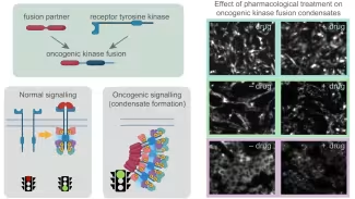
Cells rely on tightly regulated signaling networks to control growth, survival, and responses to their environment. In cancer and other diseases, these pathways become deregulated and are frequently rewired by oncogenic mutations. Our laboratory studies abnormal signaling in kinase-driven cancers, focusing on how oncogenic alterations reshape protein interaction and phospho-signaling networks.
Current projects include studies of oncogenic kinase fusions and truncations, signaling condensates and membraneless organelles, Hippo pathway signaling, receptor tyrosine kinase signaling, stress-responsive signaling systems, and related adaptive signaling pathways. We use proximity labeling, quantitative proteomics, phosphoproteomics, and functional genomics to define how signaling networks are dynamically organized and remodeled in disease states. These studies integrate proteomics, structural biology, computational approaches, and cancer models to uncover how adaptive signaling states create therapeutic vulnerabilities.
Nutrient sensing, mTOR signaling and cancer metabolism
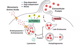
Cells continuously monitor nutrient availability and adapt their growth and metabolism accordingly. A central regulator of this process is mTORC1, a protein kinase complex that integrates nutrient and growth factor signals to coordinate anabolic and catabolic programs. Dysregulation of mTOR signaling is a hallmark of many cancers, including KRAS-driven lung and pancreatic cancers.
Our research seeks to understand how cancer cells adapt nutrient sensing and metabolic signaling pathways to survive fluctuating and nutrient-limited environments. A major focus is on defining how alternative nutrient sensing mechanisms and lysosomal signaling pathways reshape mTOR-dependent growth programs and cellular adaptation.
We previously discovered an unexpected Rag-independent pathway through which amino acids generated by lysosomal protein degradation activate mTORC1, revealing an alternative mechanism of nutrient sensing. Ongoing work aims to define the molecular basis of this pathway and determine how these signaling states support tumor growth, metabolic adaptation, and therapeutic resistance.
Dynamic organization of human cells
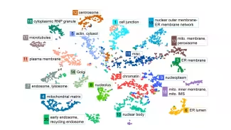
A major focus of our laboratory is developing proximity-based proteomics approaches to study how proteins are organized within living cells and how these structures change in response to stress, signaling activation, and disease. This work is particularly important for understanding membraneless organelles, dynamic cellular structures that organize biochemical reactions and establish signaling specificity without surrounding membranes.
Our group developed the Human Cell Map, a large-scale effort to define the molecular organization of human cells using proximity-dependent biotinylation and mass spectrometry. We are now extending these approaches to generate dynamic maps of cellular organization across diverse biological contexts by integrating proteomics, imaging, and advanced computational analysis.
These efforts have contributed to the development of widely used datasets, analytical frameworks, and experimental strategies that support the broader proteomics and cell biology communities.
Spatial proteomics and cell-state transitions
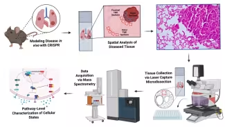
Tissues are complex ecosystems composed of many interacting cell types, and their spatial organization strongly influences disease progression and therapeutic response. Our laboratory develops spatial proteomics technologies that enable protein analysis directly within tissues while preserving spatial context and cellular organization.
These projects integrate spatial biology, functional genomics, and precision medicine approaches to understand how signaling pathways and cellular states evolve during tumor progression and therapy response, and how interactions between tumour, immune, and stromal cells shape disease adaptation.
A major current focus, in collaboration with Drs. Schramek and Campbell, is lung adenocarcinoma, where we combine spatial proteomics, in vivo CRISPR screening, and computational approaches to investigate how genetic alterations reshape tumour biology and the surrounding microenvironment. We are also developing workflows for phosphoproteomics and proteomic analysis of extremely small tissue regions and rare cell populations from both clinical samples and animal models.
Proteomics technology development and computational biology
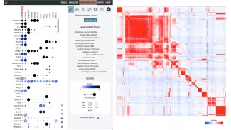
Technology innovation is deeply integrated into our research program. We develop scalable proteomics and computational approaches to study rare cells, small tissue regions, and dynamic cellular processes with increasing sensitivity and spatial resolution.
Our group has contributed to widely adopted experimental, computational, and community resource platforms for protein interaction mapping, proximity labeling, and systems-level proteomics, including tools for data visualization and functional annotation. Current efforts include low-input microproteomics workflows, spatial and proximity proteomics technologies, and machine learning approaches for integrating large-scale proteomic datasets and modeling signaling-state transitions.
Pandemic preparedness and host-pathogen biology
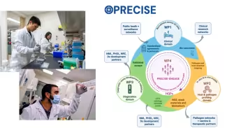
During the COVID-19 pandemic, our laboratory helped establish scalable serology and protein-based diagnostic platforms to support multiple research studies and public health initiatives across Canada. In collaboration with clinicians, immunologists, structural biologists, and national research networks, we developed assays to measure immune responses to SARS-CoV-2 infection and vaccination and helped deploy these technologies in large population studies.
Building on these efforts, we now co-lead with Dr. Johnstone PRECISE, a multidisciplinary initiative focused on pandemic preparedness, rapid diagnostic development, and emerging infectious threats. Our ongoing work includes immune profiling, blood proteomics, host-pathogen interaction studies, viral interactome mapping, and technologies for rapid characterization of immune responses and pathogen biology.
These studies integrate proteomics, immunology, virology, structural biology, and clinical research to support translational science and public health readiness while generating scalable tools and datasets that can be broadly deployed across research and healthcare settings.
We are always looking for motivated researchers to join our team.
Postdocs
Our research group is always interested in recruiting highly motivated postdoctoral fellows with a strong publication record in one of our areas of research or technology development. Please forward your CV, references and research interests to Anne-Claude Gingras.
Graduate students
Our research group is part of the Department of Molecular Genetics at the Temerty Faculty of Medicine of the University of Toronto, which has a central admission committee and a rotation system. Graduate students interested in doing a PhD in the laboratory must first be accepted in the Department of Molecular Genetics.
Summer students
Summer students are exclusively selected from successful applicants to the Research Training Center (RTC) at the Lunenfeld-Tanenbaum Research Institute. Applications are available online and need to be filled by February 28th of each year.
Notable publications
Science Signaling, 2024
Science, 2020
Science Immunology, 2020
Molecular Cell, 2018