Dr. Ian Rogers
Lunenfeld-Tanenbaum Research Institute
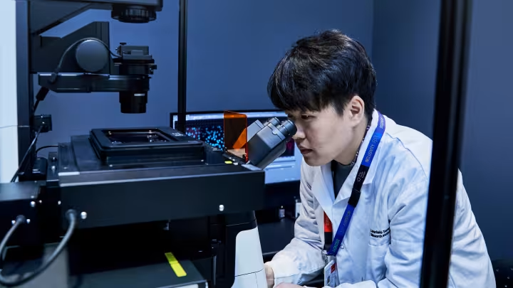
Regenerative medicine: cell therapy and bioengineered organs
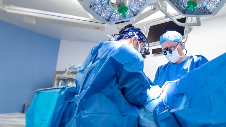
Our research harnesses the power of stem cells –the ability to self-renew and turn into specialized cells – to study embryo development and advance regenerative medicine. For many diseases, organ transplant is the only effective treatment yet donor organ shortages leave many patients without options. Advances in stem cell biology now offer the possibility of generating transplantable tissue and organs, including patient-matched grafts that could eliminate the need for life-long immune suppression.
We have three aims: 1) to use ex vivo organ perfusion to maintain organs outside the body for drug discovery and development studies, 2) to discover new treatments for kidney fibrosis and type1 diabetes using bioengineered patient-derived and functional mini-organs and 3) to bioengineer patient-matched organs from stem cells for transplantation.
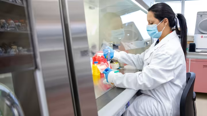
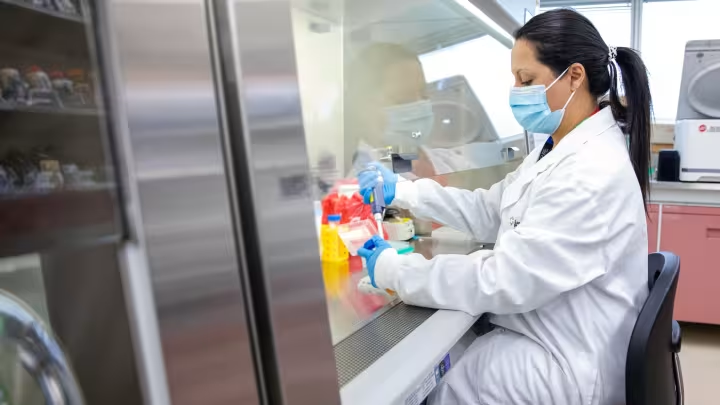
We use decellularized animal organs—cell-free scaffolds that retain native structure—as templates to guide stem cell growth and differentiation. We have showed that mouse kidney and lung scaffolds seeded with stem cells can generate partially functional organized tissues. Building on this work, we are now using larger scaffolds to develop patient-specific human pancreas and kidney for future studies and transplantation.
A key finding from our work is that the extracellular matrix, a meshwork of molecules surrounding our organs, guides stem cell differentiation into specialized organ cells. This discovery underpins our strategy for building functional organs and optimizing culture conditions. We have also developed a custom bioreactor system that sustains organs ex vivo, enabling long-term studies of tissue growth and function.
We also work with induced pluripotent stem cells (iPSCs), derived from adult cells. We contributed to a global multi-omics study characterizing iPSCs at genetic, epigenetic, and protein levels, helping advance their application in regenerative medicine.
Email: [email protected]
Room 5-1015, 25 Orde St.
Toronto, M5T 3H7
Website: Website name
Publications: PubMed
Google Scholar: Ian Rogers
ORCID: 0000-0001-8466-1225
U of T Department of Physiology: Ian Rogers
U of T Department of Obstetrics & Gynaecology: Ian Rogers
LinkedIn: Ian Rogers
- 2025–present; Translational Research Scientist, Biomedical Stream, Lunenfeld-Tanenbaum Research Institute, Sinai Health, Toronto
- 2024–present; Professor, Institute of Medical Sciences, Faculty of Medicine, University of Toronto, Toronto
- 2023–present; Professor, Departments of Obstetrics and Gynaecology, Physiology and Laboratory Medicine & Pathology, Faculty of Medicine, University of Toronto, Toronto
- 2021–present; Associate Scientist, Ajmera Transplant Center, Faculty of Medicine, University of Toronto, Toronto
- 2020–present; Founder, 3.G.O medicine, Toronto
- 2014–present; Scientist, Ontario Institute for Regenerative Medicine (formerly OSCI), Toronto
- 2014–present; Research Scientist, Banting and Best Diabetes Centre, University of Toronto, Toronto
Former appointments
- 2015–2023; Associate Professor, Departments of Obstetrics and Gynaecology, Physiology and Laboratory Medicine & Pathology, Faculty of Medicine, University of Toronto, Toronto
- 2023–2025; Scientist, Lunenfeld-Tanenbaum Research Institute, Sinai Health, Toronto
- 2007–2023; Associate Scientist, Lunenfeld-Tanenbaum Research Institute, Sinai Health, Toronto
- 2004–2022; Co-Founder and Consultant for Inception Biosciences, Mississauga, ON, Canada. Now part of Cooper Scientific
- 2003–2015; Assistant Professor, Obstetrics and Gynaecology, Faculty of Medicine, University of Toronto, Toronto
- Post-doctoral fellowship with Dr. R. Casper, Department of Obstetrics and Gynecology, University of Toronto, Toronto; 1999–2002
- PhD, Embryology, University of Toronto, Toronto; 1994–1999
- MSc, Immunology, University of Toronto, Toronto; 1984–1987
Engineering organs: kidney, lung, pancreas
Organ repair or regeneration rely on interactions between cells and the extracellular matrix (ECM). Building on three technologies; induced pluripotent stem cells (iPSCs), ex vivo organ perfusion (EVOP), and organ decellularization, we have developed a system to study cell-ECM interactions during tissue regeneration in a 3D organ culture model. The ECM is a complex structure composed of structural proteins, proteoglycans, glycosaminoglycans, and embedded growth factors. These growth factors are critical for tissue organization, as their unique combinations provide a distinct “home address” that guides multipotent cells to differentiate into the appropriate cell types for that tissue. We are leveraging this property to engineer new organs.
Our method for decellularization removes cellular material while preserving the ECM’s protein composition. We have shown that early organ progenitor cells can interact with acellular ECM to differentiate and form organized, functional organs. Importantly, the evolutionary conservation of ECM allows us to substitute animal cells with human cells, resulting in humanized organs. These humanized tissues are used to model human disease. Unlike organoids, our humanized organs are vascularized, have advanced organization, and reach a more mature functional state.
Investigating the role of circulating immune cells on kidney fibrosis-progression and treatment
Kidney fibrosis is a major contributor to kidney failure and represents the end stage of chronic kidney disease, which can arise from multiple causes. As we age, the incidence of kidney disease increases, and the body's ability to repair damage shifts from true regeneration to fibrosis (scarring).
Fibrosis is driven by complex cellular behaviors, including altered cell proliferation, differentiation, and migration, and is strongly influenced by the immune system. All organs are affected by both tissue-resident and circulating immune cells. Circulating immune cells interact systemically, while resident immune cells are uniquely adapted to the specific organ in which they reside. Distinguishing the roles of these two immune cell populations in vivo is challenging, as there are no simple methods to separate them.
By isolating the kidney and maintaining it ex vivo, we eliminate the influence of circulating immune cells. Using established methods to induce renal fibrosis, we can compare disease initiation and progression in both ex vivo and in vivo environments to determine the specific contribution, if any, of circulating immune cells. Importantly, the ex vivo organ perfusion system also enables the controlled reintroduction of defined immune cell populations, allowing us to directly test their role in the development and progression of renal fibrosis.
Determining the mechanism of action of candidate drugs to treat type 1 diabetes
Type 1 diabetes affects millions of people globally. Current treatment regimens require constant blood glucose monitoring and insulin injections. Improvements now include an insulin pump capable of monitoring glucose and dispensing insulin automatically. Recent results from a clinical trial demonstrated that stem cell-derived islets can be successfully transplanted and remain functional for one year. The recipients did not require exogenous insulin. This is very promising. Despite the success, unmatched stem cell-derived islets are still susceptible to both rejection due to being unmatched and the autoimmune reaction that caused the diabetes initially.
We are working on an alternative treatment that uses drugs to promote endogenous ß-cell expansion. Understanding its mechanism of action is important for further drug development. Isolated islet culture is difficult to sustain and not practical for these studies and in vivo mouse models are limited due to the inaccessibility of the pancreas and the inability to follow changes in real time. We have combined our EVOP system with fluorescent reporters for α and ß cells. This way we can observe in real time ß cell proliferation and/or α to ß cell transdifferentiation. Function via a Glucose Stimulated Insulin Secretion (GSIS) test can be done at the same time. This system can be used as a drug screening/ discovery platform.
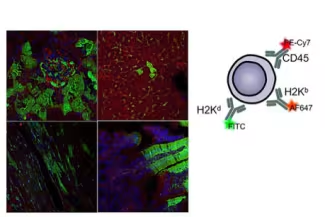
Immune tolerance during kidney transplantation: Enhancing trogocytosis in kidney transplantation in order to promote tolerance
Trogocytosis is a unique method of cell-cell communication that results in the transfer of intact, functional membrane proteins between cells without the transfer of any DNA or RNA. There is evidence that trogocytosis based transfer occurs at the immune synapse that is formed during immune based interactions. We have shown that during bone marrow transplant the recipient HLA proteins (that define ‘self’) are transferred to the donor cells thus giving them protection from rejection. Given that gene and protein expression is highly regulated, the reason cells would capture and present proteins from other cells is still elusive.
In my lab we use both in vitro and in vivo experiments to develop a more comprehensive understanding of the role for trogocytosis in immune system function. By determining the underlying mechanism we aim to uncover the signals that control trogocytosis. This should help us to promote tolerance in other transplant situations such as kidney transplants.
Investigation of the role of the uterus on embryo development
The uterus plays both mechanical and biological roles in fetal development. However, current studies on mammalian fetal development are largely limited to in vivo animal models, making it difficult to isolate and study the effects of specific external factors. To address this challenge and enable more precise control over developmental influences, we have developed an ex vivo organ perfusion system for the pregnant uterus. This allows us to regulate the composition of the perfusate and introduce specific hormones, proteins, peptides, or drugs to assess their direct impact on development.
Hormones also have a role in regulating blood flow rate and pressure, uterus muscle tension and other mechanical influences that can be controlled through perfusion pump settings, vasodilators, oxygen supply and medium composition. Therefore, the uterus ex vivo perfusion system allows us to deconstruct different contributing factors. This will be important for deciphering the intricacies of the role of the uterus in pregnancy.
We are always looking for motivated researchers to join our team.
Postdocs
Our research group is always interested in recruiting highly motivated postdoctoral fellows with a strong publication record in tissue engineering and/or embryo development Please forward your CV, references and research interests to Dr. Ian Rogers. Email: [email protected]
Graduate students
Our research group is part of the Departments of Physiology and Laboratory Medicine & Pathology, and the Institute of Medical Sciences at the University of Toronto. It is suggested that a supervisor be identified before admission to the graduate program. Graduate students interested in doing a PhD with my laboratory/group should first contact Dr. Ian Rogers ([email protected]), directly.
Summer students / Project students
Students should contact Dr. Ian Rogers ([email protected]), directly.
Notable publications
Scientific Reports, 2023
Biomaterials Science, 2022
Cells, 2022
NPJ Regenerative Medicine, 2020
Join our team
Visit our job board to see research positions.